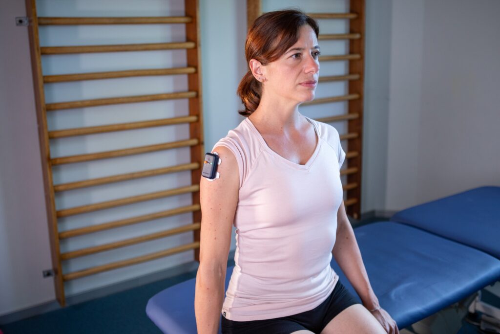
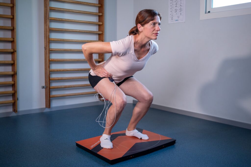
Surface electromyography (surface EMG or sEMG) is a non-invasive technique that records the electrical signals generated by muscles during contraction, providing insights that extend far beyond visual observation or palpation. This technology has become an essential tool in physical therapy, rehabilitation, sports performance, and ergonomics, offering valuable insights for diagnosis, training optimization, and patient education.
In this article, we explore what surface EMG is, its benefits, limitations, applications, and how to prepare accurate assessments. With portable sensors like the K-Myo from Kinvent, practitioners can perform high-precision muscle analysis anywhere, from clinics to sports fields, empowering data-driven decisions in rehabilitation, performance, and research.
CONTENTS
1- What is a surface EMG (electromyography)?
2- Advantages and potentials of surface EMG
3- Limitations of surface EMG
4- Applications of surface EMG
5- How are EMG signals detected?
6- Components of the EMG signal
7- Key factors influencing EMG signal quality and interpretation
8- Strategies for effective electrode placement in surface EMG
9- How to prepare an assessment with an EMG device
10- FAQ
11- Conclusion
12- References
1- What is a surface EMG?
Surface electromyography (sEMG) is a specialized technique for recording, analyzing, and interpreting myoelectric signals, the tiny electrical impulses produced when muscle fibers are activated. These signals reflect the physiological changes in muscle cell membranes during contraction.

Unlike traditional clinical EMG in neurology, which often uses needles and focuses on artificially induced muscle responses in static conditions, sEMG measures voluntary muscle activity in real-life situations. This makes it particularly valuable for assessing posture, dynamic movement, work-related tasks, sports performance, and rehabilitation exercises.
By translating these electrical patterns into a visual graph, sEMG allows professionals to observe how muscles activate, coordinate, and relax. When combined with other biomechanical tools, such as inertial measurement units (IMUs) or force plates, it provides a comprehensive picture of muscle function and neuromuscular control.
2- Advantages and potentials of surface EMG
Surface EMG stands out as a safe, non-invasive, and accurate method for analyzing muscle activity. Unlike visual observation or palpation, it delivers objective, quantifiable data on how muscles work, both at rest and during movement.
Key advantages include:
- Comprehensive muscle insight: sEMG reveals activation patterns, timing, and coordination that are invisible to the naked eye.
- Real-time visual feedback: Graphical displays enable professionals and patients to instantly understand muscle function.
- Integration with other sensors: When paired with grip dynamometers, traction dynamometers, handheld dynamometers, or force plates, sEMG provides a complete view of biomechanics.
- Supports evidence-based practice: Data can be shared with peers, researchers, and insurers to justify treatment choices.
- Enhances patient engagement: Seeing their muscle activity helps patients better follow rehabilitation guidance.

By answering questions such as “Is the target muscle activating correctly?” or “Is there a compensatory pattern?”, sEMG becomes a valuable sensor for optimizing training, rehabilitation, and performance.
💡 To learn more, watch the webinar “EMG Biofeedback in Physical Therapy: Elevate Your Assessments with Muscle Activation Insights” and discover how to make the most of surface EMG to analyze muscle activation, optimize rehabilitation, and enhance performance.
3- Limitations of surface EMG
While surface EMG is a powerful assessment tool, it has certain limitations that users must consider:
- Limited monitoring scope: It can be challenging to record multiple muscle sites simultaneously, especially for complex movements.
- Muscle substitution: Different muscles may produce similar movements, making it harder to isolate the target muscle.
- Crosstalk: Signals from nearby muscles can interfere with recordings, reducing specificity.
- Standardization issues: Variations in electrode placement between sessions or practitioners can affect data consistency.
- Interpretation limits: sEMG measures electrical activity, not muscle strength; results require proper normalization for comparison.
Understanding these constraints and applying consistent, precise measurement techniques ensures that sEMG delivers reliable and meaningful insights.
4- Applications of surface EMG
Surface EMG has evolved from a physiological and biomechanical research tool to a versatile solution used in healthcare, sports, and industry.
Physical therapy & Rehabilitation
- Monitor muscle activation post-surgery or after neurological injury.
- Track patient progress and adapt therapy plans.
- Provide biofeedback to improve motor control.
👉 Practical example: Preventing quadriceps inhibition after knee surgery
In post-surgical knee rehabilitation, surface EMG is particularly valuable for addressing inhibition of the vastus medialis oblique (VMO), a common issue that delays functional recovery. With real-time visual biofeedback, patients can immediately see whether the muscle is activating correctly. Combined with tempo training (controlled rhythm of contraction and relaxation), sEMG helps improve VMO recruitment, restore balanced quadriceps activation, and accelerate the return to walking and sports activities.
Sports Science & Performance
- Analyze muscle recruitment during training.
- Identify inefficiencies or compensations in movement.
- Personalize programs to boost performance and reduce injury risk.
Ergonomics & Workplace Health
- Evaluate muscle strain during occupational tasks.
- Support injury prevention strategies.
- Optimize equipment and workstation design.
Research & Biomechanics
- Study muscle coordination, fatigue, and force generation.
- Combine with force plates and motion capture for complete biomechanical profiling.
From rehabilitation clinics to elite sports training facilities, sEMG provides the objective data needed to optimize movement, recovery, and performance.
💡Spotlight on our neuromuscular sensor K-Myo: Bringing sEMG to life
When discussing surface electromyography (sEMG), having precise, real-time data is key. That’s where Kinvent’s K-Myo comes in. This non-invasive EMG sensor lets clinicians, therapists, and researchers capture muscle activation, coordination, fatigue, and timing effortlessly.
Whether you’re tracking rehabilitation progress, optimizing sports performance, or analyzing movement patterns, K-Myo provides visual biofeedback and measurable insights that make neuromuscular assessment intuitive and actionable. Compact, wireless, and easy to integrate with the Kinvent App, it brings EMG directly into your practice, helping you measure muscle and improve movement, one session at a time.
5- How are EMG signals detected?
Surface EMG works by detecting the tiny electrical changes that occur when muscle fibers are activated. When a nerve impulse reaches a muscle, it triggers a rapid shift in the muscle fiber’s membrane potential, known as depolarization, followed by repolarization as the fiber resets.
This electrical activity creates a potential difference that can be measured using two surface electrodes placed over the muscle. As the action potential moves along the fiber, the voltage changes between the electrodes, producing a bipolar signal that reflects the combined activity of many fibers within a motor unit.
Accurate detection depends on:
- Precise electrode placement
- High-quality amplification to capture subtle signals in the microvolt range
- Noise reduction to avoid interference from nearby muscles (crosstalk) or external sources
Through this process, sEMG translates invisible muscle activity into clear, measurable data, ready for clinical, sports, or research analysis.
6- Components of the EMG signal
Superposition of Motor Unit Action Potentials
The electrical activity detected at the electrode site is the superposition of Motor Unit Action Potentials (MUAPs) from all active motor units. This overlay produces a bipolar signal with a symmetric distribution of positive and negative amplitudes, averaging to zero. This combined signal is called an interference pattern, providing a snapshot of muscular electrical activity during contractions.
Motor Unit Recruitment and Firing Frequency
The intensity and density of the EMG signal are primarily influenced by motor unit recruitment and firing frequency.
- As additional motor units are recruited, the signal’s magnitude increases, reflecting higher muscle force output.
- Firing frequency: the rate at which a motor unit is activated also affects force generation.
It is important to note that surface EMG recordings are filtered by the skin and connective tissue, which slightly alters the original signal characteristics. Despite this, sEMG broadly reflects the recruitment and firing patterns of motor units, offering valuable insights into muscle behavior and performance.
7- Key factors influencing EMG signal quality and interpretation
For surface EMG to deliver reliable results, several factors must be controlled:
- Tissue Characteristics: Subcutaneous fat, hydration level, muscle fiber type, and blood flow can all alter signal amplitude and frequency.
- Physiological Crosstalk: Nearby muscles may generate signals that interfere with the target muscle’s activity.
- Electrode Muscle Geometry: Muscle length changes and joint movement can affect electrode distance and alignment.
- External Noise: Power lines, electronic devices, or poor grounding can contaminate the signal.
- Electrodes & Amplifiers: Type, size, placement, inter-electrode distance, and device quality directly influence data accuracy.
By addressing these variables through proper setup and testing, practitioners can ensure that their sEMG recordings truly reflect the target muscle’s activity.
8- Strategies for effective electrode placement in surface EMG
Correct electrode placement is essential for capturing accurate and reproducible sEMG data. The electrodes serve as the primary sensors for detecting muscle activity, and their positioning directly impacts the quality of the signal. Although literature on this topic is limited, several principles have been identified to optimize sEMG recordings.
- First, it is important to position electrodes as close as possible to the target muscle. Reducing the tissue layer between the electrodes and the muscle fibers minimizes signal loss and interference, ensuring a more accurate recording.
- Equally critical is the alignment of the electrodes with the muscle fibers. Electrodes placed parallel to the fiber orientation maximize sensitivity and specificity, while those positioned perpendicular to the fibers may compromise selectivity and reduce the clarity of the recorded signal.
- Another key consideration is avoiding the motor end plate region. Placement over the innervation zone can lead to lower recorded amplitudes due to differential amplification. Ideally, electrodes should be positioned along the midline of the muscle belly, midway between the innervation zone and the tendon, to capture a strong and representative signal.
- Using clear anatomical landmarks further enhances reproducibility across sessions and among different practitioners. Consistent placement ensures that recordings remain precise over time, which is essential for longitudinal studies, clinical assessments, or performance monitoring.
- Practical considerations also play a role: electrodes should not obstruct movement or vision and should avoid skin creases, bony prominences, or other areas that could compromise placement or comfort.
- Finally, minimizing interference from adjacent muscles is crucial. Choosing the appropriate electrode size and spacing helps limit crosstalk, isolating the desired muscle activity and improving the reliability of the measurements. By carefully considering these factors, clinicians, researchers, and sports professionals can significantly enhance the accuracy and reproducibility of their sEMG recordings.
Following these guidelines greatly improves sEMG data quality and makes comparisons between sessions more reliable.
9- How to prepare an assessment with an EMG device
A well-prepared setup is crucial for obtaining high-quality surface EMG data. Follow these steps:
- Electrode placement: Use anatomical landmarks, place electrodes parallel to muscle fibers, close together, and select the smallest appropriate size.
- Skin preparation: Remove hair, clean the area with an abrasive material and alcohol to lower skin impedance.
- Baseline check: Verify noise level, zero offset, and stability during joint movement before starting measurements.
- Signal validation: Perform static test contractions to confirm that the target muscle is being recorded accurately.
- Measurement consistency: Use a flexible tape to measure inter-electrode distance and keep placement consistent across sessions (see SENIAM guidelines).
This preparation ensures your sEMG results are reliable, reproducible, and ready for meaningful interpretation.
10- FAQ
What is the difference between EMG and surface EMG?
Traditional EMG often uses needle electrodes to record activity from deep muscles in clinical neurology. Surface EMG (sEMG) uses electrodes placed on the skin to measure voluntary muscle activity, making it non-invasive and suitable for movement analysis.
Can surface EMG be used in sports?
Yes. It’s widely used in sports science to analyze muscle recruitment, detect compensations, optimize training programs, and reduce injury risk.
Is surface EMG painful?
No. It’s entirely non-invasive and involves only the placement of electrodes on the skin.
How accurate is surface EMG?
Accuracy depends on factors such as electrode placement, skin preparation, and signal quality. Following best practices ensures reliable, reproducible results.
11- Conclusion
Surface EMG has transformed the way we assess and understand muscle function. By providing real-time, objective data on muscle activation, coordination, and relaxation, it empowers clinicians, coaches, and researchers to make precise, evidence-based decisions. From rehabilitation and performance enhancement to ergonomic assessments, its applications are broad and impactful.
With portable, high-precision devices like the K-Myo from Kinvent, you can bring advanced muscle analysis to any setting, maximizing accessibility and efficiency. Whether in a clinic, gym, or research lab, sEMG enables you to track progress, optimize interventions, and engage patients or athletes in their performance journey.
12- References
- BASMAJAN J.V., DE LUCA CJ. Muscles Alive: Their Functions Revealed by Electromyography. Williams & Wilkins, 5th Edition, 1985.
- MERLETTI, R., & PARKER, P. (2004). Electromyography: physiology, engineering, and non-invasive applications. John Wiley & Sons.
- CRAM JR, STEGER JC. EMG scanning and the diagnosis of chronic pain. Biofeedback Self Regul. 1983;8:229–241.
- FARINA, D., MERLETTI, R., & ENOKA, R. M. (2014). The extraction of neural strategies from the surface EMG. Journal of Applied Physiology, 117(5), 491-501.
- ENOKA, R. M. (2002). Neuromechanics of human movement. Human Kinetics.
- DE LUCA, C. J. (1997). The use of surface electromyography in biomechanics. Journal of Applied Biomechanics, 13(2), 135-163.
- FRIDLUND AJ, CACIOPPO JT. Guidelines for human electromyographic research. Psychophysiology. 1986 Sep;23(5):567-89. doi: 10.1111/j.1469-8986.1986.tb00676.x. PMID: 3809364.
- KASMAN G, CRAM JR, WOLF S. Clinical Applications in Surface Electromyography. Gaithersburg, MD: Aspen; 1997
- TAYLOR W. Dynamic EMG biofeedback in assessment and treatment using a neuromuscular reeducation model. In: Cram JR, ed. Clinical EMG for Surface Recordings, II.